A cikk orvosi szakértője

Új kiadványok
Sötétedés a röntgenfelvételeken felnőtteknél és gyermekeknél
Utolsó ellenőrzés: 29.06.2025

Minden iLive-tartalmat orvosi szempontból felülvizsgáltak vagy tényszerűen ellenőriznek, hogy a lehető legtöbb tényszerű pontosságot biztosítsák.
Szigorú beszerzési iránymutatásunk van, és csak a jó hírű média oldalakhoz, az akadémiai kutatóintézetekhez és, ha lehetséges, orvosilag felülvizsgált tanulmányokhoz kapcsolódik. Ne feledje, hogy a zárójelben ([1], [2] stb.) Szereplő számok ezekre a tanulmányokra kattintható linkek.
Ha úgy érzi, hogy a tartalom bármely pontatlan, elavult vagy más módon megkérdőjelezhető, jelölje ki, és nyomja meg a Ctrl + Enter billentyűt.
Gyakran előfordul, hogy a diagnosztikai intézkedések részeként az orvos röntgenfelvételt ír elő a betegnek. Ezt az eljárást a szövetek és szervek károsodásának, valamint más vizsgálatok eredményeinek tisztázására végzik. A röntgenkép megfejtését egy szakképzett radiológus végzi, aki a kapott információkat elküldi a kezelőorvosnak. De egy átlagos beteg számára az ilyen "dekódolás" gyakran érthetetlen. Tehát, ha bármilyen sötétedést észlelnek a röntgenfelvételen, sok beteg feleslegesen aggódni kezd, mert nem ismeri a helyzet lényegét. Pánikba kell esni, ha a kép foltokat vagy sötétedést tartalmaz, és mit jelent ez?
Mit jelent a röntgenfelvételen megjelenő áramszünet?
Amikor a betegeknek közlik a röntgenfelvételen látható elsötétülés jelenlétét, sokan szorongni kezdenek, és rosszindulatú daganatot feltételeznek. Valójában a daganat némileg elsötétedik, de ez csak egy a tünet számos oka közül. Ezért ne ijedjen meg azonnal: fontos, hogy a lehető legtöbb információt szerezzen erről a jelenségről, és megismerkedjen a röntgenfelvételeken megjelenő elsötétülés minden lehetséges tényezőjével.
És az okok a következők lehetnek:
- A röntgengép nem megfelelő kezelése, gyenge minőségű film használata, nem megfelelő képelőhívási eljárás.
- Idegen testek jelenléte a szervekben és szövetekben.
- Korábbi sebészeti műtétek nyomai (hegek).
- Gyulladásos gócok.
- Helminth felhalmozódások, paraziták.
- Törésnyomok és egyéb csontsérülések.
- Folyadékok jelenléte.
- Jóindulatú és rosszindulatú daganatok, áttétek.
Ha az orvos röntgenfelvételen sötétedést észlel, további diagnosztikára lehet szükség. Ez a betegség okainak és árnyalatainak tisztázásához szükséges. A kezelést csak az összes vizsgálat elvégzése után lehet felírni. Ezenkívül figyelembe veszik a beteg panaszait, klinikai tüneteit és általános jólétét.
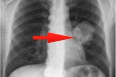
Hogyan nézhet ki a tüdő áramszünete röntgenfelvételen?
A tüdő sötétedését az orvos a következő mutatók alapján értékeli:
- A sötétedés lokalizációja fent, lent, a tüdő középső részében van. Ezenkívül a sötétedés a tüdő külső, középső vagy belső lebenyében is elhelyezkedhet.
- A sötétedés mértéke fontos a kóros folyamat területének felméréséhez.
- A sötétedés intenzitása segít meghatározni a fókusz sűrűségét (közepes, gyenge és hangsúlyos).
- A körvonalak általános jellemzői: a szegélyek laposak, szaggatottak stb. Gyakran ez a mutató teszi lehetővé egy adott betegség gyanúját.
Ezek a legfontosabb, de nem minden jel, amire az orvos a megfejtés és a diagnózis során figyel. Ezenkívül figyelembe kell venni a röntgenfelvételen látható sötétedés típusát és alakját is:
- A lebenykék sötétedésének éles határai vannak, egyfajta homorúság vagy konvexitás. Gyulladásos vagy destruktív folyamat jele lehet. A tüdő középső-alsó zónájában lokalizálódó lebenykék sötétedésének daganatképződésre utalhat.
- A tüdő röntgenfelvételén látható gócos sötétedés egy kis (kb. 10 mm-es) folt, amely gyulladásos folyamatra, érrendszeri patológiára, perifériás rák, tuberkulózis vagy tüdőinfarktus kialakulására utal. Ha a gócokat a beteg fejfájás, köhögés és mellkasi nyomás panaszaival együtt észlelik, akkor bronchopneumóniára lehet gyanakodni.
- A meghatározatlan alakú sötétedések általában nem intenzívek és nem rendelkeznek egyértelmű konfigurációval. Ilyen helyzetben a diagnózis felállításához további diagnosztikát írnak elő - különösen mágneses rezonancia vagy komputertomográfia. Leggyakrabban az elmosódott foltok mellhártyagyulladás, tüdőgyulladás vagy valamilyen daganatos folyamat jelei.
- A folyadék sötétedése a tüdőödéma biztos jele. A magas érnyomás miatt nedvesség gyűlhet fel, ami fokozott érpermeabilitással jár. Ilyen helyzetben a tüdőfunkció kifejezett károsodása áll fenn.
- A szegmentális sötétedés egy háromszöghöz hasonlít. Ez rosszindulatú betegségekben, tuberkulózisban, tüdőgyulladásban stb. fordul elő. Ilyen helyzetben fontos, hogy az orvos megfelelő képzettséggel rendelkezzen - mind a diagnózis, mind a kezelés hozzáértő előírása tekintetében.
- A fokális sötétedés egyetlen, legfeljebb 10 mm méretű folt. Ez a jel gyakran tüdőgyulladásra, tuberkulózisra, cisztás és gennyes tömegekre utal.
Egy megfelelő szakember soha nem fog diagnózist felállítani kizárólag a röntgenfelvételeken látható sötét foltok típusa és lokalizációja alapján. Általában teljes körű diagnózisra van szükség, beleértve a laboratóriumi vizsgálatokat is.
Amikor az orvos kóros tünetek kombinációjával szembesül, további diagnózisra van szükség. Továbbá, ha sötétedést észlelnek, az orvosnak meg kell különböztetnie a betegséget, és meg kell válaszolnia a következő kérdéseket:
- Specifikusnak bizonyult a folt vagy sem (tuberkulózis)?
- A sötétedés gyulladásos reakcióra utal?
- Lehet, hogy rosszindulatú folyamatról van szó?
- Van-e bizonyíték bármilyen ritka (nem gyakori) patológiára?
Jobb tüdő sötétedése röntgenfelvételen.
Fontos megérteni, hogy a tüdő bármilyen sötétedése, akár jobbra, akár balra, nem diagnózis, hanem csak a betegség egyik jele. Hogy milyen betegségről beszélünk, az a teljes diagnosztikai komplexum után válik világossá. Ennek eredményeként az orvos összehasonlítja az összes eredményt és tünetet, és csak ezután fog végleges diagnózist készíteni.
A tüdőpatológiákat többnyire a tüdőszövet különböző megvastagodásának gócai kísérik. Ez a szerv egyes területein a légáramlás romlása vagy teljes elzáródása következtében történik. Az ilyen pecsétek a röntgenfelvételen sötétedésnek tűnnek.
A túlnyomórészt jobb oldalon megjelenő apró, fókuszsötétedések tüdőbetegség kialakulására utalhatnak. Egyetlen kép vizsgálatával nem lehet egyértelműen megválaszolni a probléma okára és eredetére vonatkozó kérdéseket. Ezért kiegészítő diagnosztikai típusokat kell kijelölni - például CT-t, MRI-t, vagy ugyanazt a röntgenfelvételt, de más szögekből. Ezenkívül a laboratóriumban vizeletet, vért, köpetváladékot stb. is vizsgálnak.
Ha a röntgenfelvételen apró sötétedések láthatók olyan tünetekkel egyidejűleg, mint láz, gyengeség, fejfájás, köhögés, mellkasi fájdalom, akkor tüdőgyulladás (bronchopneumonia) gyanúja merülhet fel.
Ha a laboratóriumi vizsgálatok nem mutatnak nyilvánvaló változásokat a vérben, az lehetővé teszi számunkra, hogy fontolóra vegyük a gócos tuberkulózis jelenlétét. Ilyen helyzetben a beteg étvágytalanságra, fáradtságérzésre, száraz köhögésre, mellkasi fájdalomra panaszkodik. A gyanú kizárására vagy megerősítésére megfelelő vizsgálatokat írnak elő.
A tüdőinfarktusban a legtöbb betegnél az alsó végtagok tromboflebitise, szív- és érrendszeri rendellenességek, oldalirányú mellkasi fájdalom és néha hemoptysis fordul elő.
A tüdőrák egy rosszindulatú betegség, amely gyakrabban alakul ki a jobb tüdőben. A felső lebenyek gyakrabban érintettek, mint az alsók. Ezért a tüdő felső részének röntgenfelvételen látható sötétedése riasztónak kell lennie, és további gondos diagnózis, beleértve a differenciáldiagnózist is, okot kell, hogy adjon: ezt a jelenséget meg kell különböztetni a tuberkulózistól.
Ezek a leggyakoribb patológiák, amelyeket a röntgenfelvételen áramszünetek formájában rögzítenek. Számos más, kevésbé gyakori patológia is létezik, és ezek kialakulásának valószínűségét is figyelembe kell venni.
Sötétedés a tüdőben egy gyermek röntgenfelvételén
A tüdő sötétedésének kimutatása gyermekgyógyászati betegeknél speciális megközelítést igényel. A kép értelmezésének a lehető legrészletesebbnek kell lennie, az összes kóros elváltozás teljes jellemzőjével.
- A bal vagy jobb oldalon megnagyobbodott tüdőgyökerek leggyakrabban hörghurutra vagy tüdőgyulladásra utalnak.
- A tüdő bal vagy jobb oldalán található elmélyült érrendszeri mintázat a légzőrendszerben károsodott vérkeringésre, szív- és érrendszeri problémákra utal, de a hörghurut, a tüdőgyulladás vagy az onkopatológia kezdeti szakaszának jele is lehet.
- A fibrózis (fibrotikus szövet) jelenléte a légzőrendszer korábbi műtéti beavatkozásának vagy traumájának eredménye.
- A fokális árnyékok jelenléte az érrendszeri mintázat egyidejű fokozódásával a tüdőgyulladás tipikus képe.
Az észlelt sötétedés számos különböző betegségre utalhat. Ezért ne diagnosztizálja saját maga a gyermeket. Fontos a diagnózis folytatása. Például az orvos az alábbi vizsgálatokat írhatja elő:
- Diaskin teszt (előnyben részesített) vagy Mantoux teszt;
- Köpet elemzés;
- A tüdő CT-vizsgálata;
- Bronchoszkópia, tracheobronchoszkópia;
- Általános vérvizsgálat, biokémiai vérvizsgálat, onkomarker teszt.
Bizonyos vizsgálatok szükségességét egyénileg határozzák meg.
A csont sötétedése a röntgenfelvételen
A csont- és ízületi rendszer röntgenfelvétele az egyik gyakori diagnosztikai módszer, amely segít a diagnózis felállításában, a szövődmények azonosításában és a további kezelés meghatározásában. Először is, ilyen vizsgálatot akkor végeznek, ha törések, csonttörések, ficamok és szubluxációk, szalagsérülések gyanúja merül fel. Lehetőség van másodlagos csont- és ízületi rendellenességek, degenerációs folyamatok stb. kimutatására is.
Csonttörés esetén a sérült terület lineáris világosodása figyelhető meg a megmaradt szerkezeti sötétedés hátterében. A törésvonal nem minden esetben látható.
Csontritkulás esetén a csontszövetben a kalciumsók sűrűsége csökken, amit a röntgenfelvételeken sötétedő területek formájában észlelnek. Ha a rendellenesség kifejezett, a szerkezet jól átengedi a röntgensugarakat, ami nyilvánvaló sötét foltok megjelenéséhez vezet.
Az asszimilált csonthártyagyulladás a kalciumlerakódások és az alatta lévő csontok ízületi tapadását mutatja, amelyet meg kell különböztetni a zúzódásos törés utáni túlzott csontkeményedéstől.
A fasciák, inak, szalagok sérülése vérömlenyek kialakulását okozza, amelyekben kalcium sók rakódnak le, így ez a folyamat a képen sötétedéssel látható. Az ilyen patológia okai lehetnek trauma, fizikai túlterhelés stb.
A bordákon a röntgenfelvételeken látható sötétedés, akárcsak más csontokon, a törés utáni csontkallusz kialakulása során jelenik meg. Ebben az esetben a kallusz a csontgyógyulás során képződő kötőszöveti terület. Radiológiailag a regenerációs folyamat a következőképpen néz ki:
- Néhány hét elteltével gyengén intenzív, muffle alakú sötétedés jelenik meg a csontos kerület mentén;
- A kimaradás intenzitása fokozatosan növekszik;
- A csontkallusz kialakulásának befejezése után a kerület kifejezett sötétedését határozzák meg, és a töredékek között csontgerendák jelennek meg.
Az orrmelléküregek sötétedése röntgenfelvételen.
Mennyire veszélyes lehet az orr röntgenfelvételen való elsötétülése? Ezt a következtetést gyakran hangoztatják a fül-orr-gégészeti szervek különböző patológiáinak diagnosztizálásakor. Egyszerűen fogalmazva, a sötétedés leggyakrabban gyulladásos reakciót jelez az egyik vagy másik szakaszban (arcüreg), váladékozás megjelenésével. A röntgenvizsgálatot gyakran ajánlják arcüreggyulladásban, frontitiszben és arcüreggyulladásban szenvedő betegeknek.
A röntgenfelvételen látható a maxilláris és frontális arcüreg, valamint a rácsos labirintus. A sötétedés intenzitása lehetővé teszi a betegség stádiumának és elhanyagolásának felmérését. A kifejezett árnyékok a gennyes váladék erős felhalmozódását jelzik - azaz a kórokozó flóra aktív szaporodását. A maxilláris és frontális gyulladás kórokozói leggyakrabban pneumokokkuszok és streptokokkuszok, amelyek különösen az elhúzódó nátha hátterében válnak aktívvá, ha a kezelést nem végezték el, vagy írástudatlanok voltak. A gyulladásos reakció a nyálkahártya duzzanatát okozza, ami blokkolja a felhalmozódott váladék kiválasztását, ami további tényezővé válik a mikrobák fokozott szaporodásában.
A röntgenfelvételen a maxilláris arcüreg sötétedése a nyálkahártya szöveteinek megvastagodásával járhat, ami a következők eredményeként történik:
- Akut gyulladásos folyamat esetén;
- Az allergiás folyamatról;
- Elhúzódó krónikus gyulladás esetén.
A problémát azonban nem csak gyulladás okozhatja – például a röntgenfelvételen sötét homloküreg cisztát jelenthet, amely a képen jól látható. További okok lehetnek az orrmandula és a polip, amelyek különösen hajlamosak az orrfolyásra, és idővel arcüreggyulladáshoz vezethetnek.
A patológia fejlődési stádiumának felmérésére az orrmelléküregek röntgenfelvételét írják elő. Például, ha a folyamat kellően elhanyagolt, akkor a röntgenfelvételen részösszegű vagy teljes sötétedés formájában jelentkezhet.
A különféle orrmelléküreg-váladékok jellegzetes röntgenjele a „tej a pohárban”. Ez a tünet a folyadék azon tulajdonsága miatt jelentkezett, hogy mindig vízszintes helyzetben van, függetlenül a beteg helyzetétől. A röntgenfelvételen látható sötétedés ebben az esetben lehet egyoldali vagy kétoldali.
Amikor egy arcüreggyulladás gyanújával kezelt beteg képét értelmezzük, felfigyelünk a folyadék jelenlétére, amely sötét háttér előtt, világos kontúrral jelenik meg. Erős gyulladásos folyamat esetén az orr felett sötétedés észlelhető, és ha árnyékok vannak jelen egyszerre több üregben is, akkor nem arcüreggyulladásról, hanem frontitiszről beszélünk. Mivel az orrmelléküregek röntgenfelvételen való sötétedése nem mindig jelent gyulladás jelenlétét, az orvos kontrasztanyagos röntgenvizsgálatot is előírhat. Ez a cisztás és daganatos daganatok meghatározásához szükséges, amelyek egyértelműen lekerekített kontúr formájában jelennek meg.
Az orrfolyás akkor következik be, ha idegen test kerül az orrüregbe.
Sötétedés a fogászati röntgenfelvételeken
A röntgenfelvételeket széles körben alkalmazzák az orvosi és ortopéd fogászatban, a szájsebészetben, a traumatológiában, valamint cisztás és daganatos képződmények kimutatásában. Ez a diagnosztikai módszer segít meghatározni a fogak állapotát anélkül, hogy megnyitnák azokat, tisztázza a gyökércsatornák számát. A röntgenfelvétel különösen nélkülözhetetlen a fogászati implantáció előtt: a kép lehetővé teszi a csontszövet térfogatának felmérését és szerkezetének vizsgálatát, ami az implantátum helyes és minőségi elhelyezéséhez szükséges.
A fogszuvasodás enyhe stádiumai súlyos zománckárosodás nélkül nem láthatók röntgenfelvételeken. A fogszuvasodás csak közepes vagy mély stádiumban, illetve szövődmények kialakulásakor észlelhető:
- A fogszuvasodás röntgenfelvételeken korlátozott sötétedésnek tűnik, csökkent sűrűséggel;
- A bonyolult fogszuvasodás a fog alakjának és anatómiai szerkezetének zavaraként jelenik meg, számos granulómával és fogcsomóval.
A röntgenfelvételeken a pulpitist a fog középső vagy alsó részének sötétedése jelzi. Súlyos betegség esetén a képen fogcsomók láthatók - különböző mennyiségű tömörödött üregek a gyökérkezelés területén.
A fogciszták sötét gócokként jelennek meg, amelyek a foggyökér területén helyezkednek el. Az ilyen gócoknak egyenletes határaik vannak, és nem egyesülnek a környező szövetekkel. Bizonyos esetekben a ciszták egyszerre két fogat is érinthetnek.
A periodontitis egy gennyes folyamat a gyökérzónában, amely röntgenfelvételen egy kis zsák formájában sötétedő elszíneződésnek tűnik.
A szív sötétedése a röntgenfelvételen
A mellkasi szervek radiológiai vizsgálata során azonosítható egy ovális alakú szívárnyék, amely a bal oldalon a ferde vonal mentén helyezkedik el. A szívizom sűrű, sötétedő, homogén szerkezetű, tiszta és egyenletes körvonalakkal és ív alakú konfigurációval rendelkezik. Az ívek mindegyike egy adott szívkamrát mutat, és kiegyenesítve a szívizom patológiájára utalnak.
A szív közvetlen elsötétülése mellett a röntgenfelvételek a következőket mutathatják:
- Érrendszeri vagy billentyűmeszesedések;
- A tüdőmintázat változásai;
- A perikardiális nyáktömlő tágulása.
A szív árnyékának ilyen variációi vannak:
- Jobbkezes pozicionálás;
- A pleurális üregbe való eltolódással (effúzió miatt);
- Egy daganat vagy rekeszizomsérv által elmozdítva;
- A tüdőzsugorodás miatti elmozdulással.
A röntgensötétedést a perikardiális membrán gyulladásos folyamataiban észlelik (folyadék jelenléte a szív körül, a perikardiális lemezek között), kalciumlerakódással az erek falán (koszorúér-kalcinózis).
A szívröntgen kétféleképpen végezhető: standard kontrasztanyag nélkül vagy kontrasztanyaggal a bal pitvar szélének jobb megvilágítása érdekében.
A röntgenfelvételeken látható sötétedés veszélyes tüdő- és egyéb patológiákra, valamint alacsony minőségű filmre utalhat. Ezért ilyen helyzetben ne essen pánikba, mert a röntgen csak egy a diagnosztikai módszerek közül, és az orvos soha nem fog végleges diagnózist felállítani pusztán a kép alapján.
Általánosságban elmondható, hogy a röntgenfelvételen látható sötétedés fehér foltnak tűnik (mivel negatív képet használnak), de eredetét számos ok okozhatja. A helyzet tisztázása érdekében számos további vizsgálatot kell előírni, valamint szükség esetén röntgenfelvételt egy másik vetületben.