A cikk orvosi szakértője
Új kiadványok
Sötétedés a röntgenfelvételeken felnőtteknél és gyermekeknél
Utolsó ellenőrzés: 07.06.2024

Minden iLive-tartalmat orvosi szempontból felülvizsgáltak vagy tényszerűen ellenőriznek, hogy a lehető legtöbb tényszerű pontosságot biztosítsák.
Szigorú beszerzési iránymutatásunk van, és csak a jó hírű média oldalakhoz, az akadémiai kutatóintézetekhez és, ha lehetséges, orvosilag felülvizsgált tanulmányokhoz kapcsolódik. Ne feledje, hogy a zárójelben ([1], [2] stb.) Szereplő számok ezekre a tanulmányokra kattintható linkek.
Ha úgy érzi, hogy a tartalom bármely pontatlan, elavult vagy más módon megkérdőjelezhető, jelölje ki, és nyomja meg a Ctrl + Enter billentyűt.

Gyakran, a diagnosztikai intézkedések részeként az orvos felírja a beteg radiográfiáját. Ezt az eljárást a patológiák, a szövetek és a szervek károsodásának kimutatására, valamint más vizsgálatok eredményeinek tisztázására hajtják végre. A röntgenkép megfejtése egy speciális radiológussal foglalkozik, aki ezután elküldi a kapott információkat a kezelőorvosnak. De az átlagos beteg esetében az ilyen "dekódolás" gyakran érthetetlen. Tehát, ha valamilyen sötétítést észlelnek a röntgenfelvételen, sok beteg feleslegesen aggódik, mert nem ismerik a helyzet lényegét. Pánikba kell esnünk, ha a kép foltokat vagy sötétítést tartalmaz, és mit jelent ez?
Mit jelent a röntgenban lévő áramszünet?
Amikor a betegeknek elmondják az áramszünet jelenlétéről a röntgenfelvételen, sokan szorongnak, feltételezve a rosszindulatú daganatokat. Valójában egy daganat sötétítést mutat, de ez csak egy a tünet sok oka. Ezért ne félj azonnal: Fontos, hogy minél több információt szerezzünk erről a jelenségről, és megismerkedjünk a röntgenfelvételek sötétedésének előfordulásának minden lehetséges tényezőjével.
És az okok a következők lehetnek:
- A röntgengép nem megfelelő működése, alacsony minőségű film használata, nem megfelelő képfejlesztési eljárás.
- Az idegen testek jelenléte szervekben és szövetekben.
- A korábbi műtéti műveletek (hegek) nyomai.
- Gyulladásos fókuszok.
- Helmint felhalmozódás, paraziták.
- Törési jelek és más csont sérülések.
- A folyadékok jelenléte.
- Jóindulatú és rosszindulatú daganatok, metasztázisok.
Ha az orvos a röntgen sötétítését észlel, további diagnosztikára lehet szükség. Ez szükséges a betegség okainak és árnyalatainak tisztázására. A kezelés csak az összes vizsgálat befejezése után írható be. Ezenkívül a beteg panaszait, a klinikai tüneteket, az általános jólétet figyelembe veszik.
Hogyan nézhet ki a tüdőben lévő áramszünet egy röntgenfelvételen?
A tüdő sötétedését az orvos ezen mutatók értékelik:
- A sötétítés lokalizációja alatt, lent, a tüdő középső részén. Ezenkívül a sötétítés a tüdő külső, középső vagy belső lebenyében helyezkedhet el.
- A sötétítés mérete fontos a patológiás folyamat területének felméréséhez.
- A sötétítés intenzitása segít meghatározni a fókusz sűrűségét (közepes, gyenge és kiejtett).
- A körvonalak általános jellemzői: A határok laposak, egyenesek stb.
Ezek a legfontosabbak, de nem minden olyan jel, amelyre az orvos figyel a megfejtés és a diagnózis során. Ezenkívül figyelembe kell venni a röntgen sötétítésének típust és alakját:
- A lobularis sötétítésnek egyértelmű határai vannak, egyfajta konkavitás vagy domborúság. Ez lehet a gyulladásos vagy pusztító folyamat jele. A lobuláris sötétedés lokalizációja a tüdő középső lower zónájában jelezheti a tumorképződést.
- A tüdőben a röntgenfelvétel fókuszos sötétedése egy kicsi (kb. 10 mm) folt, amely gyulladásos folyamatot, érrendszeri patológiát, vagy perifériás rák, tuberkulózis vagy tüdőfarktus kialakulását jelzi. Ha a fókuszokat észlelik a beteg fejfájdalmával, köhögésével és nyomásával a mellkasban, akkor a bronchopneumonia gyanúja lehet.
- A meghatározatlan formájú sötétségek általában nem intenzívek, és nem rendelkeznek egyértelmű konfigurációval. Az ilyen helyzet diagnosztizálásához további diagnosztikát írnak elő - különösen a mágneses rezonancia vagy a számítógépes tomográfia. A leggyakrabban elmosódott foltok a pleurisy, a tüdőgyulladás vagy néhány daganat folyamata jele.
- A folyadék sötétítése a tüdőödéma biztos jele. A nedvesség a magas érrendszeri nyomás miatt összegyűjtheti a megnövekedett érrendszeri permeabilitást. Ilyen helyzetben a pulmonális funkció kiemelt károsodása van.
- A szegmentális sötétítés hasonló a háromszöghez. Ez rosszindulatú betegségekben, tuberkulózisban, tüdőgyulladásban és így tovább fordul elő. Ilyen helyzetben fontos, hogy az orvos elegendő képesítéssel rendelkezik - mind a diagnózis, mind a kezelés kompetens receptje szempontjából.
- A fókuszos sötétítés egyetlen, legfeljebb 10 mm méretű. Ez a jel gyakran a tüdőgyulladás, a tuberkulózis, a cisztás és a zűrzavar tömegét jelzi.
A megfelelő szakember soha nem fog diagnózist végezni kizárólag a röntgenfelvételek típusa és lokalizációja alapján. Általában teljes átfogó diagnózisra van szükség, beleértve a laboratóriumi vizsgálatokat is.
Ha az orvos a patológiás tünetek kombinációjával szembesül, akkor a további diagnózisnak szükségszerűen be kell tartania. Sőt, amikor sötétítést észlelnek, az orvosnak meg kell különböztetnie a betegséget, és válaszolnia kell az ilyen kérdésekre:
- A folt specifikus vagy sem (tuberkulózis)?
- Van-e a sötétítésnek a gyulladásos reakció bizonyítéka?
- Lehet, hogy rosszindulatú folyamat?
- Van-e bizonyíték valamilyen ritka (ritka) patológiáról?
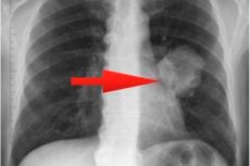
A jobb tüdő sötétedése a röntgenfelvételen.
Fontos megérteni, hogy a tüdő bármilyen sötétedése, jobb vagy bal, nem diagnózis, hanem csak a betegség egyik jele. Milyen betegségről beszélünk, a diagnosztika teljes komplexe után világossá válik. Ennek eredményeként az orvos összehasonlítja az összes eredményt és tünetet, és csak akkor végez végső diagnózist.
A pulmonális patológiákat többnyire a tüdőszövet vastagodási fókuszai kíséri. Ez történik a légáramlás romlása vagy teljes elzáródása eredményeként a szerv egyes területein. A röntgenkép ilyen pecsétjei sötétedésnek tűnik.
A kis fókuszos sötétítések, elsősorban a jobb oldalon, jelezhetik a tüdőbetegség kialakulását. Csak egy kép megvizsgálásával nem lehet egyértelműen megválaszolni a probléma okával és eredetével kapcsolatos kérdéseket. Ezért ki kell nevezni a diagnosztika kiegészítő típusait - például CT, MRI vagy ugyanazt a radiográfiát, de más szögekből kell végrehajtani. Ezenkívül a vizeletet, a vért, a köpet szekréciókat stb. A laboratóriumban is megvizsgálják.
Ha a röntgenfelvételek kicsi sötétítését egyszerre észlelik olyan tünetekkel, mint a láz, gyengeség, fejfájás, köhögés, mellkasi fájdalom, akkor gyanítható a tüdőgyulladás (bronchopneumonia).
Ha a laboratóriumi vizsgálatok nem mutatnak nyilvánvaló változásokat a vérben, akkor ez lehetővé teszi számunkra, hogy gondolkodjunk a fókuszos tuberkulózis jelenlétéről. Ilyen helyzetben a beteg panaszokat ad a rossz étvágyról, a fáradtság érzéséről, a száraz köhögésről, a mellkasi fájdalomról. A gyanú kizárásához vagy megerősítéséhez megfelelő teszteket írnak elő.
Pulmonális infarktus esetén a legtöbb betegnél az alsó végtagok tromboflebitiszei, a kardiovaszkuláris rendellenességek, az oldalsó mellkasi fájdalom és néha hemoptízis találhatók.
A tüdőrák rosszindulatú betegség, amely gyakrabban alakul ki a jobb tüdőben. A felső lebenyeket gyakrabban érinti, mint az alsó lebenyek. Ezért a tüdő felső részének elsötétülésének riasztónak kell lennie, és oka lehet a további gondos diagnózis oka, ideértve a differenciáldiagnózist is: ezt a jelenséget meg kell különböztetni a tuberkulózistól.
Ezek a leggyakoribb patológiák, amelyeket a röntgenképen feljegyzés formájában rögzítenek. Van azonban számos más kevésbé gyakori patológiája, és fejlődésük valószínűségét szintén figyelembe kell venni.
Sötétedés a tüdőben egy gyermek röntgenfelvételén
A pulmonális sötétítés kimutatása gyermekkori betegekben speciális megközelítést igényel. A kép értelmezésének a lehető leg részletesebbnek kell lennie, az összes patológiás változás teljes jellemzőivel.
- A megnövekedett tüdőgyökerek a bal vagy a jobb oldalon leggyakrabban bronchitist vagy tüdőgyulladást jeleznek.
- A bal vagy a jobb oldalon lévő tüdő mélyített érrendszeri mintázata azt jelzi, hogy a légzőrendszerben a vérkeringés, a kardiovaszkuláris problémák, de a bronchitis, a tüdőgyulladás vagy az onkopatológia kezdeti stádiumának jele lehet.
- A fibrózis (fibrotikus szövet) jelenléte a korábbi műtét vagy a légzőrendszer trauma eredménye.
- A fókuszos árnyékok jelenléte, az érrendszeri mintázat egyidejű javításával, a tüdőgyulladás tipikus képe.
Az észlelt elsötétítés számos különféle betegséget jelezhet. Ezért nem szabad egyedül diagnosztizálnia a gyermeket. Fontos a diagnózis folytatása. Például az orvos felírhatja az ilyen típusú tanulmányokat:
- Diaskin-teszt (előnyben részesített) vagy Mantoux-teszt;
- Köpetanalízis;
- A tüdő CT vizsgálata;
- Bronchoszkópia, tracheobronchoscopia;
- Általános vérvizsgálat, biokémiai vérvizsgálat, oncomarker teszt.
Az egyes tesztek szükségességét egyéni alapon határozzuk meg.
A csonton a röntgen sötétedése
A csont- és ízületi rendszer röntgenfelvétele az egyik leggyakoribb diagnosztikai módszer, amely elősegíti a diagnózis kialakítását, a szövődmények azonosítását és a további kezelés meghatározását. Mindenekelőtt egy ilyen vizsgálatot végeznek, amikor töréseket, csonttöréseket, diszlokációkat és szubluxációkat, ligamentum sérüléseket gyanítanak. A másodlagos csont- és ízületi rendellenességek, a degenerációs folyamatok stb. Detektálása is lehetséges.
A csonttörés során a sérült terület lineáris ragyogása van, a fennmaradó szerkezeti elsötétítés hátterében. A törésvonal nem minden esetben látható.
Az osteoporosis esetén a csontszövetben a kalciumsók sűrűsége csökken, amelyet a röntgenfelvételeken a sötétítés területén kell megfigyelni. Ha a rendellenesség kiejtett jellegű, akkor a szerkezet jól továbbítja a röntgenfelvételeket, ami nyilvánvaló sötét foltok megjelenéséhez vezet.
Az asszimilált periostitis feltárja a mögöttes csonttal rendelkező kalciumlerakódások artikulációit, amelyeket meg kell különböztetni a túlzott csont kallustól egy rögzített törés után.
A fasciae, az inak, a szalagok károsodása hematómák képződését okozza, amelyben a kalciumsók lerakódnak, tehát ezt a folyamatot a kép sötétedésével látják. Az ilyen patológia okai lehetnek trauma, fizikai túlterhelés stb.
A röntgenfelvételek sötétedése, mint a többi csontnál, a csont kallusz képződése során törés után jelenik meg. Ebben az esetben a kallusz a kötőszövet olyan területe, amely a csontgyógyulás során képződik. Radiológiai szempontból a regenerációs folyamat az alábbiak szerint néz ki:
- Néhány hét elteltével egy gyengén intenzív, muzulusz alakú sötétítés jelenik meg a csontos kerület mentén;
- Az áramszünet intenzitása fokozatosan növekszik;
- A csont kallusz képződésének befejezése után meghatározzuk a kerület kiemelt elsötétülését, és a csontok megjelennek a fragmensek között.
A sinusok sötétedése a röntgenfelvételen.
Mennyire veszélyes lehet az orr sötétedése a röntgenfelvételen? Egy ilyen következtetést gyakran hangzik az ENT szervek különféle patológiáinak diagnosztizálásakor. Egyszerűen fogalmazva: a sötétítés leggyakrabban egy vagy másik szakaszban (sinus) gyulladásos reakciót jelez a kisülés megjelenésével. A röntgen vizsgálatot gyakran javasolják a szinusitis, a frontitis és a sinusitis maxillary betegek esetében.
A röntgenkép a maxillary és a frontális sinusokat, valamint a rácsos labirintust mutatja. És a sötétítés intenzitása lehetővé teszi a betegség stádiumának és elhanyagolásának felmérését. Az expresszált árnyékok azt jelzik, hogy a gennyes szekréciók erős felhalmozódnak - azaz a patogén növényvilág aktív reprodukciója. A maxillary és a frontitis kórokozó szerei leggyakrabban pneumococcusokká és streptococcikává válnak, amelyek különösen aktívak lesznek a hosszan tartó rhinitis hátterében, ha a kezelést nem végezték el, vagy írástudatlanok voltak. A gyulladásos reakció a nyálkahártya duzzanatát okozza, amely blokkolja a felhalmozódott szekréciók kiválasztását, ami további tényezővé válik a mikrobák fokozott szorzásához.
A röntgenfelvételi szinusz sötétedése kombinálható a nyálkahártya-szövetek megvastagodásával, ami ennek eredményeként történik:
- Akut gyulladásos folyamat;
- Az allergiás folyamat;
- Hosszan tartó krónikus gyulladás.
A problémát azonban nemcsak a gyulladás okozhatja - például egy sötétített frontális sinus a röntgenfelvételen cisztát jelenthet, amelyet a képen egyértelműen látnak el. További okok lehetnek az adenoidok és a polipok, amelyek különösen hajlamosak a orrfolyásra, és idővel sinusitishez vezethetnek.
A sinusok radiográfiáját a patológia fejlődésének szakaszának felmérésére írják elő. Például, ha a folyamatot kellően elhanyagolják, akkor a röntgenképen lehet, hogy szubtotalis vagy teljes sötétítés formája lehet.
A szinuszok különféle szekrécióinak jellegzetes röntgenjele a "Tej egy üvegben". Ez a tünet azért történt, mert a folyadék tulajdonsága mindig vízszintes helyzetben van, a beteg helyzetétől függetlenül. A röntgen sötétedése ebben az esetben egyoldalú vagy kétoldalú lehet.
Amikor egy olyan beteg képének megfejtése, amelyben a sinusitis maxillary gyanúja gyanúja van, felhívják a figyelmet a folyadék jelenlétére, amely egy sötét háttérrel jelenik meg, fénykontúrával. Erős gyulladásos eljárással az orr fölött sötétítést észlelnek, és ha az árnyékok egyszerre vannak jelen több üregben, akkor nem a szinuszgyulladás, hanem a frontitisről szólnak. Mivel a röntgenfelvételek sötétedése nem mindig a gyulladás jelenlétét jelenti, az orvos emellett előírhatja a kontraszt radiográfiát. Ez szükséges a cisztás és a tumor daganatok meghatározásához, amelyek egyértelműen kiejtett lekerekített kontúr formájában jelennek meg.
Az áramszünetek akkor fordulnak elő, ha egy idegen test belép az orrüregbe.
Sötétedés a fogászati röntgenfelvételeken
A radiográfiát széles körben használják az orvosi és ortopédiai fogászat, a maxillofacialis műtét, a traumatológia területén, valamint a cisztás és tumor képződmények kimutatására. Ez a diagnosztikai módszer segít meghatározni a fogak állapotát anélkül, hogy kinyitná azokat, tisztázza a gyökércsatornák számát. A röntgen különösen nélkülözhetetlen a fogászati beültetés előtt: a kép lehetővé teszi a térfogat felmérését és a csontszövet szerkezetének vizsgálatát, amely az implantátum helyes és magas színvonalú elhelyezéséhez szükséges.
A fogszuvasodás enyhe stádiumai súlyos zománckárosodás nélkül nem láthatók a röntgenfelvételeken. A caries csak közepes vagy mély stádiumban észlelhető, vagy amikor a szövődmények fejlődnek:
- A caries korlátozottan sötétedik a röntgenfelvételeken, csökkent sűrűséggel;
- A bonyolult caries a fog alakjának és anatómiai szerkezetének megszakításaként jelenik meg, számos granulómával és fogszerekkel.
A röntgenfelvételek pulpitiszét a fog középső vagy alsó részében sötétedés jelzi. Ha ez a betegség súlyos folyamata, akkor a kép fogakat mutat - különféle mennyiségű tömörített üregeket a gyökércsatorna területén.
A fogcisztáknak a foggyökér területén lokalizált sötét fókuszok jelennek meg. Az ilyen fókuszoknak még határok is vannak, és nem olvadnak a közeli szövetekkel. Egyes esetekben a ciszták egyszerre két fogakat érinthetnek.
A periodontitis egy zűrzavaros eljárás a gyökérzónában, amely a röntgenfelvételeken egy kis zsák formájában sötétedésnek tűnik.
A szíven sötétedés a röntgenben
A mellkasi szervek radiológiai vizsgálata során meg lehet határozni a szív árnyékát, amely úgy néz ki, mint egy ovális, a bal oldalon ferde vonal mentén. A miokardium sűrű sötét, homogén szerkezetű, tiszta és akár körvonalakkal és ív alakú konfigurációval ad. Az ívek mindegyike egy specifikus szívkamrát mutat, és kiegyenlítve a miokardiális patológia jelenlétéről beszélnek.
A szív közvetlen sötétedése mellett a röntgen:
- Érrendszeri vagy szelep meszesedések;
- A tüdőmintázat változásai;
- A pericardialis bursa bővítése.
A szív árnyékának variációi vannak ilyenek:
- Jobb oldali pozicionálás;
- A pleurális üregbe való elmozdulással (az effúzió miatt);
- Daganat vagy diafragmatikus sérv elhagyja;
- A tüdő összehúzódás miatti elmozdulással.
A röntgendes sötétítést a pericardialis membrán gyulladásos folyamatain (a szív körüli folyadék jelenléte, a pericardialis lemezek között) detektálják, kalcium lerakódással az erek falán (koszorúér-kalcinózis).
A szívröntgen kétféle módon végezhető: standard kontrasztmentes vagy kontrasztjával, hogy jobban megvilágítsa a bal pitvari szegélyt.
A röntgenfelvételek sötétedése jelezheti mind a veszélyes tüdő-, mind az egyéb patológiákat, valamint az alacsony fokú filmet. Ezért egy ilyen helyzetben ne pánikba esjen, mert a röntgen csak a diagnosztikai módszerek egyike, és az orvos soha nem fog végleges diagnózist végezni csak a kép alapján.
Általánosságban elmondható, hogy a röntgen sötétedése fehér foltot mutat (mivel negatív képet használnak), de eredetét az okok tömege okozhatja. A helyzet tisztázása érdekében számos további tanulmányt szükségszerűen írnak elő, valamint szükség esetén egy röntgenfelvételre egy másik vetítésben.

